Home
/ Epidural Vs Subdural Hematoma Symptoms, Subdural and Epidural Hematoma | Dallas Brain Injury Lawyers : Spinal epidural hematoma diagnosis treatment.
Epidural Vs Subdural Hematoma Symptoms, Subdural and Epidural Hematoma | Dallas Brain Injury Lawyers : Spinal epidural hematoma diagnosis treatment.
Epidural Vs Subdural Hematoma Symptoms, Subdural and Epidural Hematoma | Dallas Brain Injury Lawyers : Spinal epidural hematoma diagnosis treatment.. If you have a subdural hematoma, blood is leaking out of a torn vessel into a space below the dura mater, a membrane between the brain and the skull. The middle meningeal artery lying under the temporal bone is often torn. Treatment of hematoma depends on the location, symptoms, and the clinical situation. Symptoms include ongoing headache, confusion and drowsiness, nausea and vomiting, slurred speech and changes in vision. Epidural hematoma, subdural hematoma, subarachnoid hematoma, intracerebral hemorrhage.
The blood that leaks from the artery forms a pocket that bulges out and puts pressure on the brain. Treatment of hematoma depends on the location, symptoms, and the clinical situation. Subdural haemorrhage the meninges are the connective tissue membranes that line the skull symptoms tend to be gradually progressive. You will easily learn these differences, as well as how to distinguish a subdural from an epidural hematoma on ct and other high yield facts about each. Hematoma is suspected in patients with symptoms and signs of acute, nontraumatic spinal cord compression or sudden, unexplained lower extremity paresis, particularly if a possible cause (eg, trauma, bleeding diathesis) is.
contusion vs hematoma - DriverLayer Search Engine from 3.bp.blogspot.com A subdural hematoma is a brain injury which involves blood collecting between the brain and the outermost meningeal part of the brain (called the dura). Along with their volume, the source of bleeding, the rate of formation, localization, distribution, and other factors, this is due to heavy concomitant brain damage more frequent than with epidural hematomas; Hematoma is suspected in patients with symptoms and signs of acute, nontraumatic spinal cord compression or sudden, unexplained lower extremity paresis, particularly if a possible cause (eg, trauma, bleeding diathesis) is. Often there is loss of consciousness following a head injury, a brief regaining of consciousness, and then loss of consciousness again. There are three categories of hematoma — subdural hematoma, epidural hematoma and intracerebral (intraparenchymal) hematoma. Closed head injuries, similar to traumatic brain injuries, come from blunt trauma to the. Subdural haemorrhage the meninges are the connective tissue membranes that line the skull symptoms tend to be gradually progressive. Chronic subdural hematoma (neurosurgery clinics of nord america, vol.11, n3), w.b.saunders company, 2000, usa, 573 p.
In small subdural hematomas with mild symptoms, doctors may recommend no specific treatment other than observation.
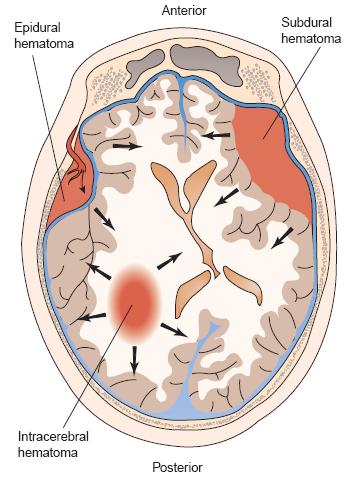
A subdural hematoma (sdh) is a type of bleeding in which a collection of blood—usually associated with a traumatic brain injury—gathers between the inner layer of the dura mater and the arachnoid mater of the meninges surrounding the brain. A subdural hematoma is a brain injury which involves blood collecting between the brain and the outermost meningeal part of the brain (called the dura). This occurs when blood vessels — usually veins — rupture between your brain and the outermost of three membrane layers that cover. Symptoms include ongoing headache, confusion and drowsiness, nausea and vomiting, slurred speech and changes in vision. Epidural hematomas occur when an artery is injured and arterial blood accumulates between the dura and the calvarium. Subdural and epidural hematomas are abnormal collections of blood within the meninges surrounding the brain. There are three categories of hematoma — subdural hematoma, epidural hematoma and intracerebral (intraparenchymal) hematoma. Blood builds up between the brain and the brain's tough outer lining. Epidural hematoma brain injuries (also referred to as extradural hemorrhages) involve blood. Epidural hematoma occurs secondary to a laceration of a vein or an artery. Spinal subdural or epidural hematoma. The term epidural hematoma refers to pooling blood (hematoma) outside the dura mater (epidural). Epidural hematoma, subdural hematoma, subarachnoid hematoma, intracerebral hemorrhage.
Do not cross suture lines because of the tight adherence of the dura to the calvarium and thus have a biconvex or elliptical appearance. More severe or dangerous subdural hematomas require surgery. Closed head injuries, similar to traumatic brain injuries, come from blunt trauma to the. There are three categories of hematoma — subdural hematoma, epidural hematoma and intracerebral (intraparenchymal) hematoma. Symptoms include ongoing headache, confusion and drowsiness, nausea and vomiting, slurred speech and changes in vision.
Epidural Hematoma, Subdural hematoma, Subarachnoid ... from i.ytimg.com Epidural hematoma is when bleeding occurs between the tough outer membrane covering the brain (dura mater) and the skull. There are three categories of hematoma — subdural hematoma, epidural hematoma and intracerebral (intraparenchymal) hematoma. Treatment of hematoma depends on the location, symptoms, and the clinical situation. An epidural hematoma typically refers to blood that acumulates between the brain and the skull after rupture of an artery secondary to trauma. Patients with an epidural hematoma can remain conscious with minimal symptoms, can become drowsy, or can progress to a coma immediately following their injury based. Here is a comparison of epidural and subdural hematomas the symptoms of subdural hematomas can vary depending on the amount of bleeding and the area of the brain that's affected. Symptoms include ongoing headache, confusion and drowsiness, nausea and vomiting, slurred speech and changes in vision. If you have a subdural hematoma, blood is leaking out of a torn vessel into a space below the dura mater, a membrane between the brain and the skull.
Chronic subdural hematoma in elderly patients:
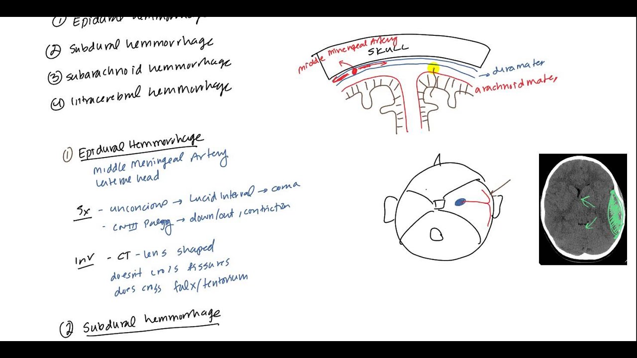
The term epidural hematoma refers to pooling blood (hematoma) outside the dura mater (epidural). Chronic subdural hematoma (neurosurgery clinics of nord america, vol.11, n3), w.b.saunders company, 2000, usa, 573 p. History and mechanism of injury. The other type of epidural hematoma is one where it occurs at the back of the brain where you fracture back here, and because it's right by the brain stem, which is the part of in a subdural hematoma the veins of your brain run in the space between the arachnoid and the dura. An epidural hematoma is usually caused by a torn artery. Patients with an epidural hematoma can remain conscious with minimal symptoms, can become drowsy, or can progress to a coma immediately following their injury based. Epidural hematoma is when bleeding occurs between the tough outer membrane covering the brain (dura mater) and the skull. Spinal subdural or epidural hematoma. Symptoms include ongoing headache, confusion and drowsiness, nausea and vomiting, slurred speech and changes in vision. Chronic subdural hematoma in elderly patients: Some may require no treatment at all while others may be deemed a. Intracranial hematomas can be epidural (above the epidural membrane), subdural (below the epidural membrane), intracerebral (within the brain tissue), or take injuries to the head seriously because of the risk of bleeding causing injuries like epidural, subdural, and intracerebral hemorrhage. Epidural hematomas occur when an artery is injured and arterial blood accumulates between the dura and the calvarium.
Significant underlying brain injury, resulting in cerebral edema. The term epidural hematoma refers to pooling blood (hematoma) outside the dura mater (epidural). Epidural hematoma occurs secondary to a laceration of a vein or an artery. Hematoma is suspected in patients with symptoms and signs of acute, nontraumatic spinal cord compression or sudden, unexplained lower extremity paresis, particularly if a possible cause (eg, trauma, bleeding diathesis) is. Symptoms of subdural hematomas are extremely variable.
Pin by Steph ls on EMT/Medical cool stuff | Subdural ... from i.pinimg.com Epidural hematoma brain injuries (also referred to as extradural hemorrhages) involve blood. Here is a comparison of epidural and subdural hematomas the symptoms of subdural hematomas can vary depending on the amount of bleeding and the area of the brain that's affected. Patients with an epidural hematoma can remain conscious with minimal symptoms, can become drowsy, or can progress to a coma immediately following their injury based. This occurs when blood vessels — usually veins — rupture between your brain and the outermost of three membrane layers that cover. Often there is loss of consciousness following a head injury, a brief regaining of consciousness, and then loss of consciousness again. Do not cross suture lines because of the tight adherence of the dura to the calvarium and thus have a biconvex or elliptical appearance. There are three categories of hematoma — subdural hematoma, epidural hematoma and intracerebral (intraparenchymal) hematoma. A subdural hematoma (sdh) is a type of bleeding in which a collection of blood—usually associated with a traumatic brain injury—gathers between the inner layer of the dura mater and the arachnoid mater of the meninges surrounding the brain.
The middle meningeal artery is classically involved.
Some may require no treatment at all while others may be deemed a. Symptoms include ongoing headache, confusion and drowsiness, nausea and vomiting, slurred speech and changes in vision. Both epidural and subdural hematomas involve bleeding outside of the brain and either outside or inside of the dura mater. Spinal subdural or epidural hematoma. A subdural hematoma (sdh) is a type of bleeding in which a collection of blood—usually associated with a traumatic brain injury—gathers between the inner layer of the dura mater and the arachnoid mater of the meninges surrounding the brain. Subdural hematoma vs epidural hematoma. The blood that leaks from the artery forms a pocket that bulges out and puts pressure on the brain. They are under low pressure. Here is a comparison of epidural and subdural hematomas the symptoms of subdural hematomas can vary depending on the amount of bleeding and the area of the brain that's affected. Spinal epidural hematoma diagnosis treatment. The features of a subdural vs an epidural hematoma differ based on ct findings, symptoms, location within the meninges, and pathophysiology. Intracranial hematomas can be epidural (above the epidural membrane), subdural (below the epidural membrane), intracerebral (within the brain tissue), or take injuries to the head seriously because of the risk of bleeding causing injuries like epidural, subdural, and intracerebral hemorrhage. Han h.j., park c.w., kim e.y., yoo c.j., kim y.b., kim w.k.
Closed head injuries, similar to traumatic brain injuries, come from blunt trauma to the epidural vs subdural hematoma. Here is a comparison of epidural and subdural hematomas the symptoms of subdural hematomas can vary depending on the amount of bleeding and the area of the brain that's affected.